Ischemic vascular diseases occur when blood flow to tissues is reduced, which can cause severe tissue damage and, in advanced cases, limb loss. Restoring blood supply is therefore a major goal of regenerative medicine. Human pluripotent stem cell (hPSC)-derived vascular organoids offer a promising source for rebuilding damaged blood vessels, but existing vascular organoids are difficult to precisely control in terms of their structure and cell composition. In addition, how transplanted human vascular cells survive and adapt inside damaged tissues remains poorly understood.
On September 25, Dr Jie Na’s group at the Aging and Regeneration Center (ARC), School of Basic Medical Sciences (BMS), Tsinghua Medicine (TM), published a research article in BMC Medicine entitled “3D bioprinted human vascular organoid sheets promote functional ischemic repair and exhibit adaptive in vivo remodeling”. The team used 3D bioprinting to build human vascular organoid sheets (hVOS) from hPSC-derived endothelial cells and smooth muscle cells. The study showed that these engineered vascular tissues can improve blood flow and promote repair in ischemic tissues, while also revealing how the transplanted cells adapt to their new environment.
Graphical abstract
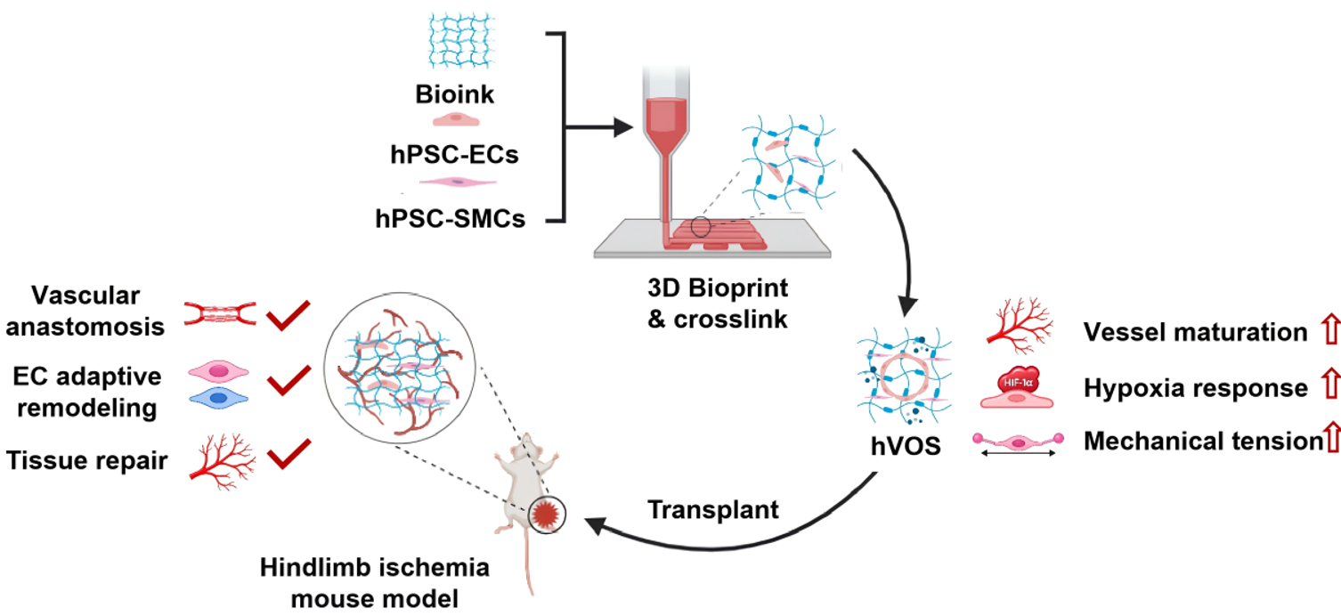
1. 3D Bioprinting Creates Human Vascular Organoid Sheets.
The researchers used 3D bioprinting to arrange hPSC-derived endothelial cells and smooth muscle cells into thin vascular organoid sheets. During culture, the cells organized themselves into vessel-like networks.
This approach provides greater control over the structure and cellular composition of the engineered vascular tissue than conventional vascular organoids. The researchers also found that the cells communicated with each other and responded to signals related to low oxygen levels and mechanical forces, which may help the vascular networks develop and mature.
2. Engineered Vascular Sheets Improve Blood Flow and Tissue Repair
The researchers then transplanted the vascular sheets into mice with reduced blood flow in the hindlimb. The transplanted human vascular cells survived for more than four weeks and grew into the ischemic tissue.
Compared with control animals, mice receiving the vascular sheets showed better recovery of blood flow and less tissue damage. Imaging also showed that vessels formed by the transplanted cells connected with the animals’ own circulation, allowing blood to flow through the newly formed vascular network.
3. Transplanted Cells Adapt to Their New Environment
The team also examined what happened to the transplanted human cells after they entered the ischemic tissue. Using single-cell analysis, they found that the vascular cells remained relatively well preserved but also changed their characteristics in response to the local environment.
At the same time, smooth muscle cells and other supporting cells showed changes associated with tissue repair and remodeling. These findings suggest that the engineered vascular tissue does not simply remain unchanged after transplantation. Instead, its cells actively respond to the damaged tissue and adapt to their new surroundings, which may help the graft integrate with the host tissue and support repair.
A New Platform for Vascular Regenerative Medicine
This study establishes a 3D-bioprinting approach for building organized human vascular tissues and demonstrates their potential to restore blood flow and promote tissue repair in ischemic conditions. Importantly, the study also provides new insight into how engineered human vascular cells survive, integrate, and adapt after transplantation.
Associate Professor Jie Na (ARC/BMS/TM, Tsinghua University; State Key Laboratory for Complex, Severe and Rare Diseases; and Beijing Key Laboratory of Intelligent Organ Biofabrication and Regenerative Repair) is the corresponding author. Xinyu Fu and Li Yan, Ph.D. candidates at Tsinghua University and Shanxi Medical University, respectively, are co-first authors. The study also received important contributions and support from Zhaosen Chen, Xinyao Zhou, Kengyuan Qu, Peiliang Wang, Bohan Dou, Dezhi Zhou, Fengzhi Zhang, Zihao Zou, Guiying Li, Taoxia Wang, Liliang Ouyang, and other collaborators from Tsinghua University, Shanxi Medical University, The First Hospital of Tsinghua University, Beijing Tsinghua Changgung Hospital, and the Affiliated Hospital of Hebei Engineering University.
This research was supported by the National Key R&D Program of China, the National Natural Science Foundation of China (NSFC), State Key Laboratory of Complex, Severe, and Rare Diseases, funding from SXMU-Tsinghua Collaborative Center for Frontier Medicine, 2026 Handan-Tsinghua Collaborative Project, the Beijing Natural Science Foundation, Hebei Provincial Key Science Technology Support Program and CPSD-Chen Jianghe Scientific Research Seed Fund.
Editors: Li Han, John Paul Grima