In a “miniature habitat” furnished with ramps, tree hollows and shelters, mice burrow into holes, climb steps and explore freely. They wear a wireless mesoscope on their heads that weighs only 1 gram excluding the battery, allowing researchers to simultaneously record the activity of thousands of neurons in their cerebral cortex.
Why make a microscope that can “run” alongside a mouse? How the brain coordinates the activity of tens of thousands of neurons across different regions during activities ranging from exploring unfamiliar surroundings to interacting and competing with peers has long been a question that neuroscientists hope to answer. In the past, microscopes able to resolve individual neurons over a large area usually required the animal’s head to be fixed or the device to be connected by cables, whereas devices that let animals move freely struggled to combine a large field of view with high imaging precision.
Developing a microscope that can “see wide and see clearly” while remaining “light to wear and free of tethers” is a challenge that Academician Qionghai Dai’s team in the Department of Automation at Tsinghua University has long sought to overcome.
On 19 October 2026, the Tsinghua team, in collaboration with Professor Pu Dai’s team at the Chinese PLA General Hospital, published a research paper online in the international journal Nature, reporting Airscope, the world’s first wireless mesoscope to exceed one megapixel.
According to the research team, the device weighs only 1 gram, more than 30% lighter than the latest head-mounted mesoscopes. It records neural activity in the mouse cerebral cortex at single-cell resolution across a 6-mm field of view and can simultaneously record more than 8,500 neurons in a single mouse, about 25 times as many as existing wireless head-mounted microscopes.
Integrating optics, electronics, artificial intelligence and other disciplines, this innovation enables scientists to observe large-scale neural activity across a wider range of natural behaviors and provides a new research tool for studying related brain disorders and for developing optical brain–computer interface technology.
“Airscope provides yet another important tool for mesoscale biology and will drive optical brain–computer interfaces toward more precise neural detection,” said Qionghai Dai. “Its applications are expected to extend from neuroscience to fields such as immunology and oncology, and to promote the industrialization of related technologies.”
Why is it so hard to see a freely moving brain clearly?
The fields of view of previous head-mounted miniature microscopes were typically smaller than 1 mm, so only a small brain area could be observed. Achieving a wider field of view and greater clarity requires acquiring and transmitting more data, which also means higher power consumption and a heavier load on the animal.
Striking a balance among field of view, imaging precision, weight and wireless transmission was the key problem the research team needed to solve.
Why observe multiple brain regions at the same time? Take picking up a cup as an example: seeing the cup, reaching out to touch it and receiving tactile feedback all require several brain regions to work together. The same holds for natural behavior: only by linking the activity of multiple brain regions can we further explore how they cooperate.
“We want to study how large numbers of cells interact with one another to give rise to the overall function of tissues and organs,” said Jiamin Wu, co-corresponding author of the paper and associate professor in the Department of Automation, Tsinghua University. “It’s a bit like the ‘sociology of cells’—what we call mesoscale biology. Take the brain: the activity of a single neuron is only one part of the picture. What matters more is understanding how large numbers of neurons connect and work together to produce complex functions.”
1 gram, wireless, high-definition: wearing a microscope on the head
Behind this breakthrough lies more than a decade of technical groundwork by the Tsinghua team.
Since 2013, under the leadership of Academician Qionghai Dai, the team has conducted sustained research on mesoscale in vivo imaging. From RUSH, the world’s first video-rate widefield mesoscopic imaging instrument, to RUSH3D, the first mesoscale in vivo 3D imaging instrument, the team has kept pushing the boundaries of our ability to observe living organisms.
As early as 2024, the team had developed a miniaturized, wearable widefield mesoscope with a 4-mm field of view, but it was still constrained by cables, making it difficult to study mammalian behavior in a fully natural state. Building on this work, the researchers continued to optimize the optical design, electronics, on-device data compression and transmission, and intelligent processing, ultimately combining a mesoscale field of view, single-cell resolution and lightweight wireless imaging. The new device complements the team’s benchtop mesoscale in vivo 3D imaging instruments and truly opens the door to mesoscale in vivo observation of multiple animals during natural activity.
“Mesoscale” can be understood as a bridge between the macroscopic and the microscopic: the aim is to cover a large expanse of tissue while still resolving the activity of individual cells within it. With this goal in mind, the research team co-designed the optics, illumination, electronics and wireless transmission as one system.
“To fit a 6-mm field of view and single-cell resolution into a 1-gram device, the optics, illumination, electronics and wireless transmission all had to be designed together,” said Yuanlong Zhang, first author of the paper and assistant professor at the School of Life Sciences and the IDG/McGovern Institute for Brain Research, Tsinghua University. “We used AI to design the lens and compressed the data about 25-fold at the sensor. That is what made the whole system light enough not to affect the mice’s natural behavior.”
For the optics, the team achieved single-cell resolution (4–6 μm) across a 6-mm field of view with only four aspheric lenses, whereas conventional head-mounted miniature microscopes typically have fields of view smaller than 1 mm. The total optical length of the lens system is also about 65% shorter than that of previous miniature microscopes. According to the team, Airscope is the smallest and lightest mesoscope published to date.
For data transmission, none of the previously published wireless systems had exceeded one megapixel. Airscope uses a 2-megapixel image sensor and compresses the data about 25-fold at the sensor, enabling megapixel-scale images to be transmitted wirelessly. The complete device (excluding the battery) weighs only 1 gram, more than 30% lighter than the latest head-mounted mesoscopes. With a magnetic quick-swap battery, researchers can replace the battery within 2 seconds and continue recording.
Experiments showed that mice wearing Airscope could climb ropes and pass through tubes, and that their distance traveled, speed, path tortuosity and other metrics did not differ significantly from those of mice without the device.
More than 8,500 neurons recorded simultaneously in a single mouse
Compared with conventional benchtop microscopes, Airscope is nearly five orders of magnitude smaller in volume, yet the quality of the neuronal signals it records remains comparable. Combined with DeepWonder, a neuron-extraction algorithm previously developed by the team (published in Nature Methods in 2023), Airscope simultaneously recorded more than 8,500 neurons across 14 cortical regions of one hemisphere in a single mouse, 25 times the number recorded by existing wireless imaging devices (typically about 300).
The team also validated the reliability of the neuronal signals through simultaneous recordings with Airscope and two-photon microscopy. Recordings from the same site could be sustained for more than 200 days.
Five mice wearing scopes at once: how does AI keep track?
Once the animals were freed from cables, the researchers faced a new problem: when several mice move about and touch one another at the same time, how can each mouse be accurately distinguished and tracked? Building on the vision foundation model SAM2, the team developed SAM2Mice, a multi-animal segmentation and tracking tool. It identifies and continuously tracks individual mice in videos and can be used in new experimental settings without re-annotation or retraining.
In a five-mouse tracking task, its multiple-object tracking accuracy (MOTA) reached 0.958, well above the 0.703 achieved by a commonly used keypoint-tracking method, and its processing speed was about 2,400 times that of expert manual annotation. Building on this, the team equipped five mice with Airscopes simultaneously in an arena 60 cm in diameter. As the mice interacted freely, more than 20,000 neurons were recorded in total, and distinct patterns of neural activity were observed during interactions. The dataset and segmentation results have been made open source.
5 square meters: observing the brain during exploration
In standard laboratory cages, mice can hardly display natural behaviors such as burrowing, long-distance exploration and complex social interaction. The team therefore built a 2.3 m × 2.3 m naturalistic habitat, laid down bedding more than 20 cm deep, and added a variety of terrain structures, including shelters, ramps, tree hollows and wooden huts.
In this habitat, the mice moved more than twice as fast as they did in their cages and displayed 16 categories of behavior, including climbing, going up and down steps, entering holes and crossing different terrain zones. The study found that changes in the terrain zone a mouse occupied were systematically linked to its cortical neural activity: from the recorded cortical activity alone, a deep-learning model could determine which zone the mouse was in, with accuracy far above chance; even when visual-cortex neurons were removed from the analysis, the model’s performance did not decline. This suggests that the zone-related information does not depend solely on the recorded visual-cortex neurons.
The team has made this “habitat–neural activity” dataset open source. According to the research team, it is the world’s first public dataset from a large naturalistic habitat that combines wide-area, single-cell-resolution cortical imaging with behavioral recordings of freely moving mice.
Who will win? Cortical activity predicts the outcome
Mice are social animals, and their groups often form a social hierarchy: who gets food first and who gains the upper hand in competition depend on more than body size. Previous studies have also suggested that social rank is not fixed, and that competitive experience may affect a mouse’s subsequent behavior. Researchers care not only about “who wins and who loses,” but also about which brain regions are active when these social behaviors occur, and how the related neural signals change as the interaction unfolds.
In the classic “tube test,” two mice meet in a narrow tube and push against each other until one of them backs out. With Airscope, the researchers could simultaneously record thousands of cortical neurons in both contestants.
The results showed that neurons associated with pushing, resisting and retreating were widely distributed across the dorsal cortex. The neural population activity of the dominant and subordinate individuals could be distinguished even before the two came into contact, and the cortical activity of the two animals was correlated above chance level.
The research team built a dual-stream Transformer deep-learning model to predict the outcome of the contest from the cortical activity of the two mice, with an accuracy of 81.8% (chance: 50%). Prediction accuracy did not decrease even when neurons associated with pushing, resisting and retreating were excluded or when a model was used to remove the influence of measured movement and behavioral variables, indicating that this cortical information cannot be explained by the measured action variables alone. A decoder trained on the standard tube test could also be transferred directly to a more naturalistic multi-loop tube network, yielding consistent results in competitions for food and for a running wheel.
Earlier studies have found that the prefrontal cortex and some thalamic nuclei are involved in social hierarchy. Because Airscope can observe a much larger area of cortex at once, it offers a new tool for exploring how competition-related activity is distributed across brain regions. Along this line, the team hopes to further investigate the neural mechanisms of social behavior and emotional states, and to provide clues for research on depression and related conditions.
From mazes to the spinal cord: expanding the frontiers of natural-behavior research
In a large binary-tree maze with 32 end points, a mouse must make five correct turns in a row to find a water reward. Airscope’s wireless design eliminates the risk of cables tangling and twisting during repeated turns, so mice can explore on their own for long periods. The study found that a latent state in cortical population activity was aligned with the moment of reward, and that this correspondence weakened after the reward location was changed.
The research team also mounted two Airscopes on a mouse, one on its head and one on its back, to image the cerebral cortex and spinal cord simultaneously during free movement, recording about 4,000 cortical neurons in one hemisphere together with population neural activity in two spinal segments.
In these simultaneous brain–spinal cord imaging experiments, the team also observed differences between the cortex and the spinal cord in vascular responses and changes in neural activity under different stimulus conditions. In a specific fear-conditioning experiment, spinal neural activity mainly preceded cortical activity in time, providing new observational data for further study of the links between activity in the brain and the spinal cord.
In addition, Airscope can be used in behavioral experiments such as swimming, tail suspension and rotating platforms, and can be synchronized with facial-expression recording and a head-mounted “first-person view” camera, linking a mouse’s facial expressions and visual perspective with its cortical activity.
From one microscope to many more studies of natural behavior
A wireless mesoscope weighing only 1 gram allows scientists to observe neural activity across large areas of cortex under conditions much closer to nature.
But “seeing” more neurons does not mean we have “understood” the brain. How do neurons in different brain regions connect with one another and work together in complex behaviors such as exploration and social interaction? Many more questions remain to be answered.
From technical breakthroughs in the laboratory to the open sharing of research results, from studies of freely moving mice to exploring the technology in other species—the Airscope story has only just begun.
The process begins with scientific questions, advances through interdisciplinary technological innovation, and ultimately expands the frontiers of science through new observational capabilities.
At Tsinghua, stories of interdisciplinary research like this continue to unfold.
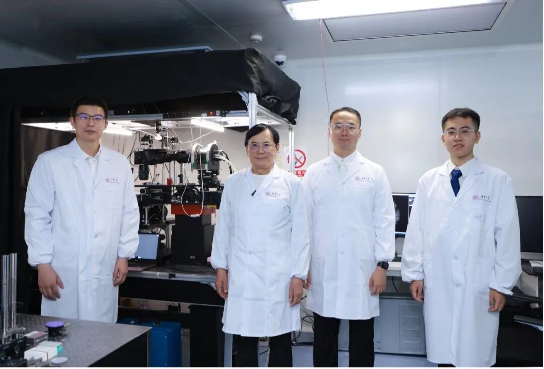
The co-first authors of the paper are Yuanlong Zhang, assistant professor at the School of Life Sciences and the IDG/McGovern Institute for Brain Research, Tsinghua University; Angran Li, a PhD student jointly trained by the College of Future Information Technology, Fudan University, and Zhongguancun Academy; Lekang Yuan, an alumnus of the 2021 master’s cohort at Tsinghua Shenzhen International Graduate School and now a PhD student in the Department of Electrical and Computer Engineering, Princeton University; Mingrui Wang, an alumnus of the 2020 doctoral cohort at Tsinghua Shenzhen International Graduate School and now an associate professor at the School of Future Technology, Northeastern University; and Dr. Weihao Zhao of the Sixth Medical Center of the Chinese PLA General Hospital. The co-corresponding authors are Associate Professor Jiamin Wu of the Department of Automation, Tsinghua University, Professor Pu Dai of the Chinese PLA General Hospital, and Professor Qionghai Dai of the Department of Automation, Tsinghua University.
This work was supported by the National Natural Science Foundation of China, the Beijing Natural Science Foundation, the Tsinghua–Peking Center for Life Sciences, and Tsinghua University’s Dushi Program.
Read the paper: Zhang, Y. et al. Cortex-wide imaging in naturally behaving mice with 1-gram wireless mesoscopes. Nature (2026). https://www.nature.com/articles/s41586-026-11293-x
Editors: Li Han, John Paul Grima